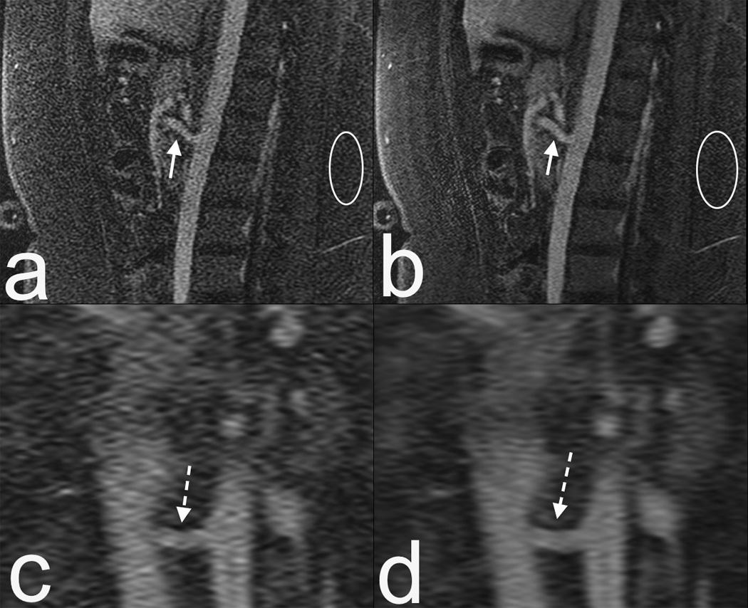
Figure 1.

16 year old female MRI at 1.5T with an 8 channel coil. (a) Sagittal contrast enhanced MRA. Note the limited SNR, particularly at the superior mesenteric artery (SMA, arrow) and noise in the subcutaneous fat (circle). (b) Compressed sensing reconstruction of same data as in (a) results in enhanced delineation of the SMA. Noise is reduced, but to a lesser degree in regions that are at the boundaries of coil sensitivities (circle). (c and d) Zoomed coronal reformats of of (a) and (b) respectively, showing enhanced delineation of the left renal artery (dashed arrow). This highlights the potential of advanced reconstruction methods.
