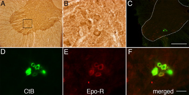
Figure 2.
Representative images of EPO-R immunostaining in C4 phrenic motor neurons. A, DAB staining revealed EPO-R expression in large, putative phrenic motor neurons (black box) and interneurons. B, Higher magnification of black bock from A. C, CtB-labeled phrenic motor neurons (green cells in C4 ventral horn). Scale bar (in C), 400 μm. D–F, EPO-R (E) is expressed in CtB-labeled phrenic motor neurons (D; see merged image in F) and the surrounding neuropil. Sections were incubated without primary or secondary antibody as negative controls. Scale bar (in F), 50 μm.

