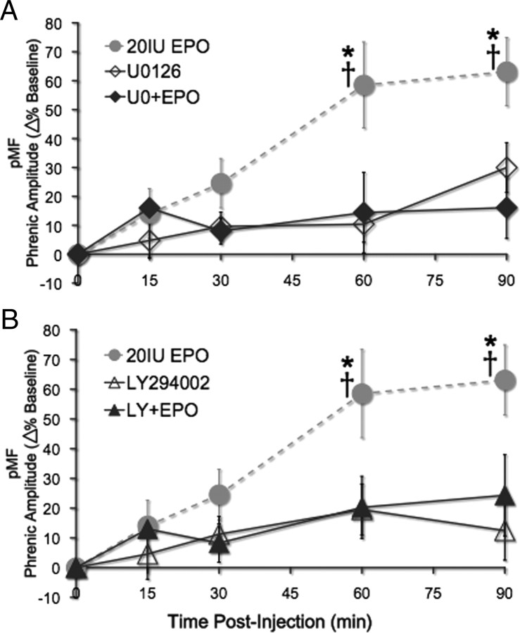
Figure 6.
EPO-induced pMF is dependent on spinal ERK and Akt activation. A, Spinal EPO elicits pMF (gray dashed line; *p < 0.001, significant difference from U0126 + EPO and U0126 alone; n = 8). Pretreatment with the MEK inhibitor U0126 abolishes EPO-induced pMF; phrenic amplitude does not increase after baseline measurement (filled diamonds; n = 5; all p > 0.05). U0126 alone had no significant effect on phrenic motor output (open diamonds; n = 6; all p > 0.05), and there was no significant difference between U0126 + EPO and U0126 alone (all p > 0.05). B, Gray dashed line shows EPO-induced pMF (* indicates significant difference from both LY294002 + EPO and LY294002 alone). After treatment with the PI3K inhibitor LY294002, EPO-induced pMF is abolished (filled triangles; n = 5; all p > 0.05). LY294002 alone had no significant effect on phrenic motor output (open triangles; n = 7; all p > 0.05), and there was no significant difference between LY294002 + EPO and LY294002 alone (all p > 0.05). All values are expressed as change in phrenic burst amplitude expressed as percentage baseline. Mean values ± 1 SEM. †p < 0.001, significantly different from baseline; *p < 0.001, significantly different from vehicle at the same time point.

