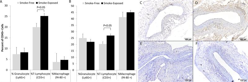
Figure 6. Leukocyte populations in the wall of experimental AAA.
Aortic tissue was harvested from mice after 6 weeks of smoke exposure (or smoke-free conditions) with aortic EP two weeks prior to harvest. Flow cytometry was performed with antibodies to CD45 (total leukocytes), CD3 (T-lymphocytes), Ly6G (neutrophils) and f4-80 (macrophages). The sub-populations of leukocytes in the spleen (A) and aorta (B) are shown as a percentage of total leukocytes. Immunohistology was performed with markers for T-cells (CD5, C and D), and B-cells (B220, E and F). The T-cells were found primarily at the outer edge of the media with some cells also found within the media. The B-cells were primarily seen in the adventitia. Although an increase in total T-cell numbers in smoke-exposed animals was evident on histology, there was no difference in the localization of these cell types based on smoke (C and E) or smoke-free conditions (D and F).

