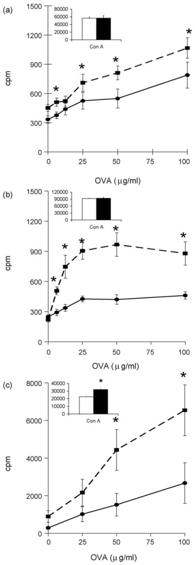
Fig. 4.

Exercise enhanced antigen-specific CD4+ T cell proliferation following i.n. vaccination. CD4+ T cells were collected from the (a) spleen (n=10/group), (b) mesenteric lymph nodes (n=5 pooled groups of 2 mice/group for each treatment) and (c) Peyer’s patches (n=5 pooled groups of 2 mice/group for each treatment) of AL (●) or AL+EX (■) mice. 1 × 10⁵ experimental CD4+ T cells were co-incubated with 5 ×10⁵ irradiated APCs from unvaccinated syngeneic mice in the presence of increasing concentrations of OVA in vitro. Insert graphs show Con A-induced CD4+ T cell proliferation from each lymphoid organ in AL (white bars) or AL+EX (black bars) mice. Data shown are mean ± SEM. Data are representative of two independent experiments. Two-way ANOVA revealed a significant effect of exercise on CD4+ T cell proliferation in response to simulation with OVA in the spleen (F 1,108 = 25.95, P < 0.001); mesenteric lymph nodes (F 1,56 = 45.43, P < 0.001); and Peyer’s patches (F 1,56 = 6.75, P = 0.012). *Post-hoc analyses using Bonferroni’s test for multiple comparisons found significant differences between AL and AL+EX groups at the designated concentration of OVA.
