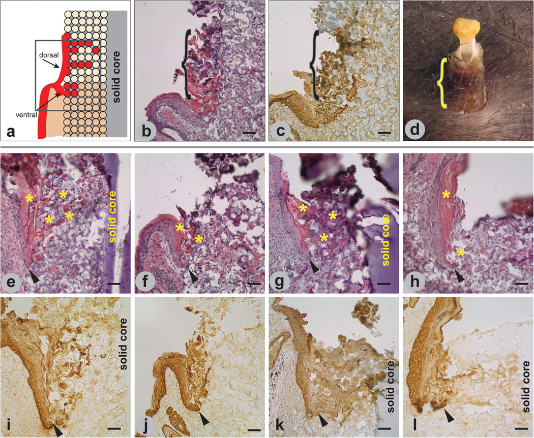
Figure 4.
Epidermal response to implanted porous/solid poly(HEMA). Illustration (a) of bifurcation of epidermis at the skin/implant interface showing keratinocytes (red) integrating into the pores from both the ventral and dorsal regions of the epidermis, with the dorsal region forming an epidermal sheath. H&E staining of implant/tissue section (b) showing cornified dorsal epidermis (pink) along and within the exterior region of a 1 1-month implant. Implant/tissue section in (c) is similar to the tissue section in (b) and immunostained with a pooled pankeratin and K14 antibody. Macro image of a 6-month implant in (d) shows external sheath formation. Brackets in (b–d) demarcate sheath. Tissue sections of 14-day (e,i), 1-month (f,j), 3-month (g,k), and 6-month (h,l) implants stained with H&E (e–h) and immunolabeled with pooled pankeratin and K14 antibody (i–l). Cornified dorsal epidermis (asterisks) is seen in (e–h). Arrowheads in (e–l) indicate tip of ventral migrating epidermis. Mag bars = 50 µm.

