
Figure 4.
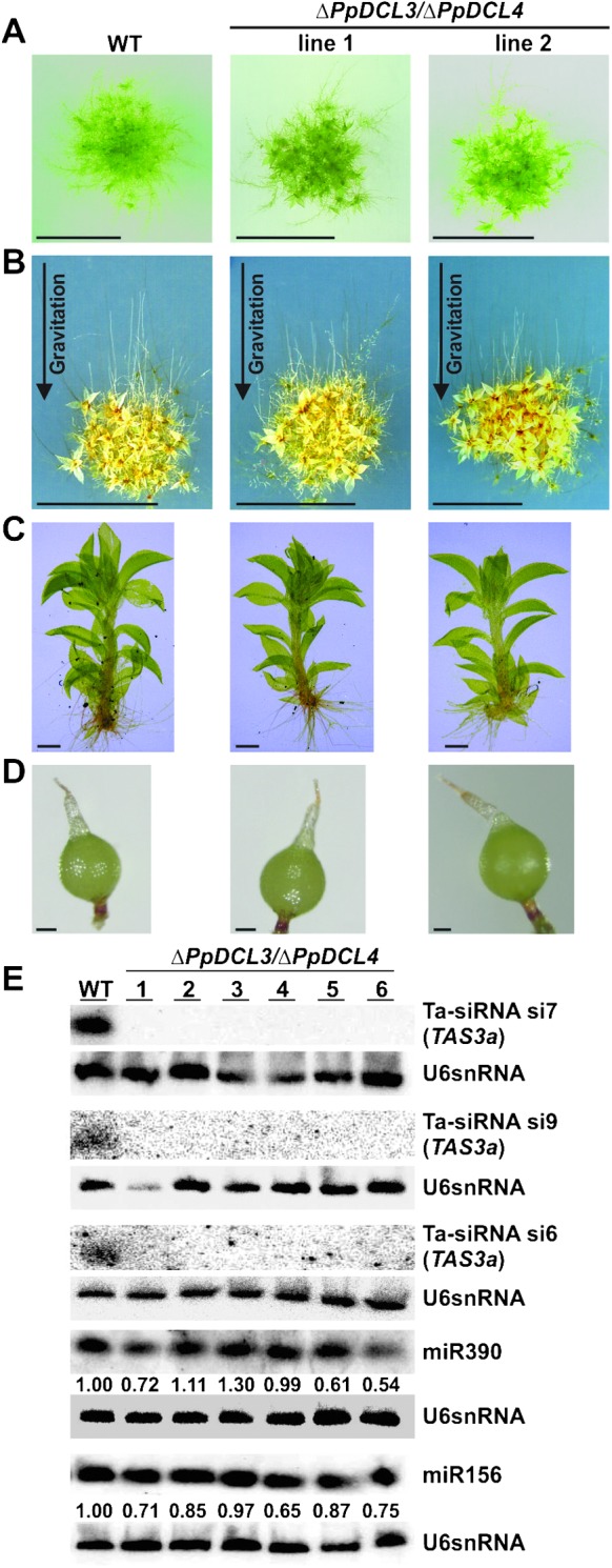
Phenotypic Analyses of ΔPpDCL3/ΔPpDCL4 Double Mutants.
(A) Equal amounts of protonema tissue from wild-type (WT) and ΔPpDCL3/ΔPpDCL4 mutants were spotted onto solid medium and developed colonies were photographed after 3 weeks. ΔPpDCL3/ΔPpDCL4 mutants show accelerated gametophore development. Scale bar: 0.5 cm.
(B) WT and ΔPpDCL3/ΔPpDCL4 mutants display normal caulonema development after 10 d growth in darkness. Scale bar: 0.5 cm.
(C) Gametophore size is similar in WT and ΔPpDCL3/ΔPpDCL4 mutants. Scale bar: 2 mm.
(D) Sporophyte formation is rescued in ΔPpDCL3/ΔPpDCL4 mutants. Scale bar: 200 μm.
(E) RNA gel blots hybridized with antisense probes of miR390, miR156, and ta-siRNAs generated from the PpTAS3a precursor in WT and ΔPpDCL3/ΔPpDCL4 double mutants. An antisense probe for U6 snRNA served as control for normalization. Numbers under blots indicate normalized miRNA expression values with respect to WT.