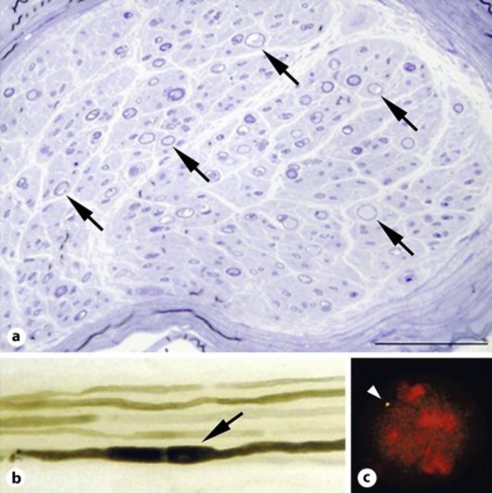
Fig. 1.
Histological and genetic results in case 1. a A cross section of the left sural nerve with toluidine blue staining showed a decrease in the myelinated fiber density. Thinly myelinated fibers were observed (arrows). Scale bar = 100 μm. b Teased fiber preparation demonstrated the characteristic tomacula formation (arrow). Other thinly myelinated fibers were observed. c Genetic analysis with the FISH method showed only one PMP22 probe signal in 97% of the white blood cells. The deletion of one copy of the PMP22 genes, compared with the presence of two copies in a normal control, supported the diagnosis of HNPP. One existing copy of the PMP22 gene is indicated (arrow head).

