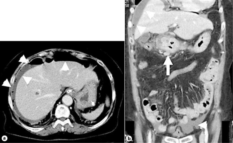
Fig. 1.
Enhanced abdominal computed tomography showed free air and ascites on the face of the liver and at the Douglas pouch (a, b; arrowheads). Levels of adipose tissue around the antrum and pylorus of the stomach revealed increase. Moreover, a thickened wall extended from the antrum of the stomach to the duodenal bulb (b) (arrow).

