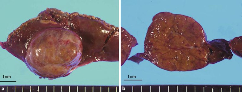
Fig. 3.
The macroscopic specimen showed a nonencapsulated solitary mass in the left lateral segment of the liver, which was 48 g and 4 × 3.5 cm in diameter (a, b). Microscopic examination showed this tumor to be composed of mature hepatocytes, ductular reaction and abnormal vessels, and revealed FNH without a central scar.

