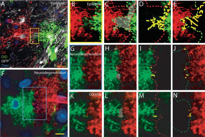
Fig. 2.
Astrocytic domain organization varies with pathology. The domain organization of protoplasmic astrocytes is lost in epileptic brains, but maintained in neurodegeneration. (a) Reactive astrocytes 1 week post-iron injection lose the domain organization. Diolistic labelling of the cortex of a GFAP-GFP mouse 1 week post-iron injection near injection site. Two adjacent GFP positive astrocytes are labeled with DiI and DiD. DAPI, blue, GFP, green, DiI, red, DiD, white. (b–e) High power of yellow box in (a). area of overlap delineated in grey, red line is border of the domain of the red cell, green line is the border of the domain of the white cell. (g–h) Yellow lines indicate the processes of the cell that pass into the domain of the adjacent cell's domain represented by the dotted line. (f) Cortical astrocytes in an Alzheimer disease model Tg2576 become reactive, but do not lose the domain organization. Diolistic labelling of cortical astrocytes in Tg2576 mouse. (g–j) High power of blue box in (f) showing limited overlap between adjacent cells. (k–n) Adjacent control astrocytes demonstrating the domain organization. Scale: (a) 20 μ m; (g–h) 10 μ m. From (22).

