
Figure 1.
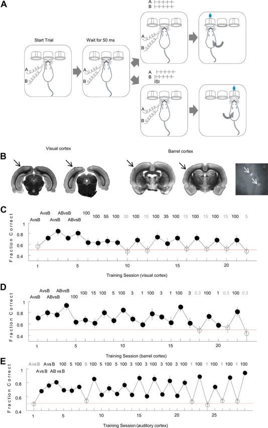
A, Experimental paradigm. Rats were implanted with two electrodes placed 1.1 mm apart, and trained to discriminate different cortical stimulation patterns. Stimuli consisted of trains of 5 pulses delivered either simultaneously (AB) or sequentially (A-ISI-B) through the two intracortical electrodes, stimulation frequency 50 Hz. Animals initiated trials by poking into a center port, which elicited a stimulus after a 50 ms delay. They were rewarded for selecting the correct reward port. B, Brain slices showing rostral (left) and caudal (right) electrode positions in visual and barrel cortices. Arrows point to the electrode positions. Cytochrome oxidase (CO) staining was used for barrel cortex. Far right panel shows CO staining of flattened barrel cortex. Arrows point to the electrode positions. C, Training history of a representative visually implanted rat. The performance on successive training sessions is plotted in chronological order. Number on top indicates ISI (ms) of the training session. Error bars indicate binomial proportion confidence interval for a 95% confidence level. Filled circles, Sessions in which the animal performed significantly above chance. Empty circles, Sessions in which performance was at chance level. D, Training history of a representative barrel cortex-implanted rat. E, Training history of a representative auditory-cortex-implanted rat (from Yang et al., 2008).