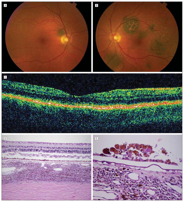
Figure 2.
Six months later, after treatment with plasmapheresis, resolution of symptoms, and return of visual acuity to 20/20 OU. A, Color fundus photograph of the right eye showing increased visibility of round, darkly pigmented tumor and resolution of subretinal fluid. B, Color fundus photograph of the left eye showing increased visibility of darkly pigmented tumors and decrease in overlying orange pigment. C, Optical coherence tomographic scan showing resolution of subretinal fluid. D, Low-power photomicrograph of the retina and choroid showing artifactual retinal detachment, diffusely thickened choroid with increased uveal melanocytic cells, areas of normal retinal pigment epithelium, and areas of hyperplastic retinal pigment epithelium (hematoxylin-eosin). E, High-power photomicrograph of hyperplastic retinal pigment epithelium, also showing some of the uveal melanocytic cells, which were mainly spindle cells with no atypia (hematoxylin-eosin).

