Figure 2.
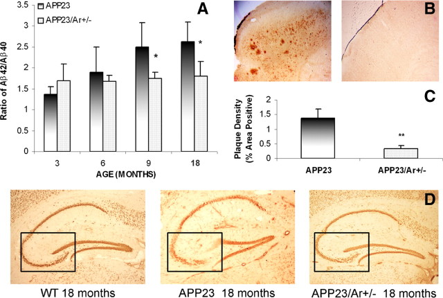
Analysis of amyloid levels, amyloid plaques and neuronal loss in the brains of 18-month-old APP23 and APP23/Ar+/− male mice. A, ELISA analysis of the reduced Aβ42/Aβ40 ratio in male APP23/Ar+/− mice compared with age-matched APP23. B, APP23 mice developed massive plaques in the cortex at the age of 18 months while small plaques were observed in the APP23/Ar+/− mice at the same age. C, The density of plaques from 19 mouse brain samples (APP23 n = 11 and APP23/Ar+/− n = 8) were analyzed. The density of plaques in APP23/Ar+/− mice were significantly less than in APP23 mice (**p < 0.001). D, Immunostaining of NeuN (1:1000) in the brain from an 18-month-old APP23 male mouse showed extensive neuronal loss in the CA3 region of the hippocampus (indicated in box) compared with the hippocampi from WT and APP23/Ar+/− male mice at the same age.

