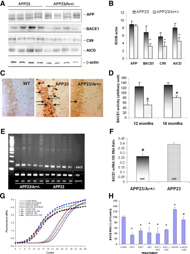
Figure 3.

Testosterone downregulates BACE1 both in vivo and in vitro. At 18 months of age, the brains from male APP23 (n = 10) and APP23/Ar+/− (n = 8) mice were harvested and prepared for Western blot, immunostaining, enzyme activity analysis and mRNA measurement. A, The Western blot of APP, BACE1, c-terminal fragment, and C99 from 18-month-old mice is shown. B, The protein levels from the Western blot are presented as the ratio of the integrated optical density value (IDV) of the protein of interest to the IDV of β-actin. C, The levels of APP c-terminal fragment in the hippocampi of 18-month-old WT, APP23 and APP23/Ar+/− mice was detected by immunohistochemistry with anti-C-terminal antibody CT-20 (1:1000) and appears as intracellular dark brown staining as indicated by the arrows. D, BACE1 enzyme activity was measured from brain homogenize of 12- and 18-month-old APP23 and APP23/Ar+/− mice. E–H, The mRNA levels of BACE1 in 18-month old APP23 and APP23/Ar+/− male mice brains were detected by RT-PCR using 18S mRNA as a control (E), and the ratios of BACE1/18S mRNA were determined by densitometric analysis (F). The effect of testosterone (T) and actinomycin D (ActD) treatments on BACE1 and 18S mRNA levels in BACE transfected 293 cells was measured by real-time RT-PCR (G); the BACE1 mRNA levels were normalized to the 18S mRNA levels and were expressed relative to the vehicle-treated cells (H). *p < 0.05 compared with APP23 mice for B, D, and F; *p < 0.05 compared with vehicle-treated cells for H.
