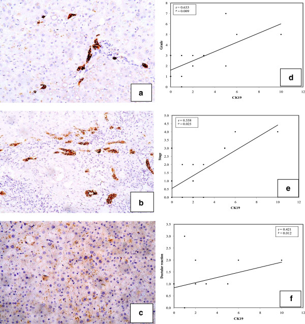
Figure 1.
Cases of hepatitis, cirrhosis and HCC stained for CK19. A case of chronic hepatitis showing (a) single scattered CK19-positive cells in the lobule (arrow) (×100). (b) A case of cirrhosis showing a higher grade of CK19-positive ductular reaction and individual positive cells in the lobule (arrow). (CK19, ×100). (c) A case of HCC showing negativity for CK19 in tumor cells. (Streptavidin-peroxidase technique, anti-CK19 monoclonal antibody, ×200).In right panels: Line graphs show significant correlations between CK19 and each (d) grade, (e) stage and (f) ductular reaction.

