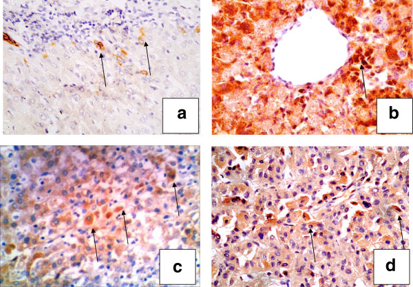
Figure 2.
Cases of hepatitis, cirrhosis and HCC stained for FGF-2. A case of chronic hepatitis showing a few positive hepatocytes in (a) periportal areas (×200), (b) the pericentral zone (×400) and (c) intermediate hepatocytes (arrow) (×200). In HCC, (d) positivity was also detected in HCC tumor cells (×200). (streptavidin-peroxidase technique, anti-FGF2 monoclonal antibody).

