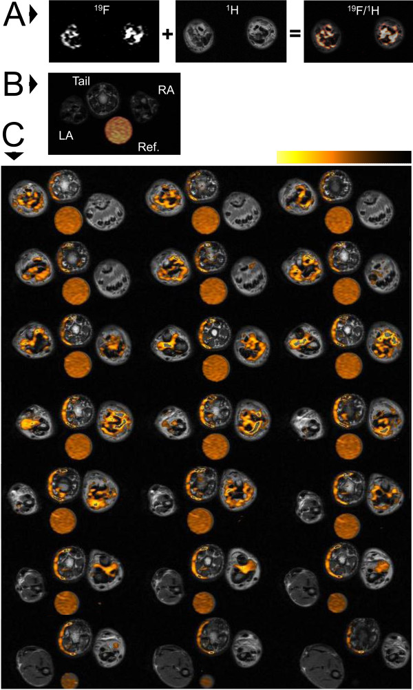
Figure 2 .
Representative 1H and 19F MRI overlays of rat ankles.A.19F signal is rendered in hot orange scale and overlayed on a grayscale 1H image in this representative slice from an arthritic rat. B. Representative slice from a naïve rat indicating the placement of right ankle (RA) left ankle (LA), tail and reference tube (Ref). C. Complete series of 19F slices obtained through the ankles of a representative arthritic rat on day 15.

