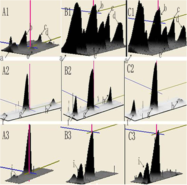
Figure 2.

3-D view of highly differentially expressed proteins. The computational analysis of the images with Image Master 2D Platinum 6.0 software allowed for the detection of protein volumes. Significant changes of more than 5-fold were found in the protein spots (spots a to i) of the magnified regions 1–3 of the corresponding 2-DE gels among the nonresponder group (A), the responder group (B), and the healthy control group (C). The differentially expressed proteins were down-regulated in the nonresponders compared with the responders and healthy controls.
