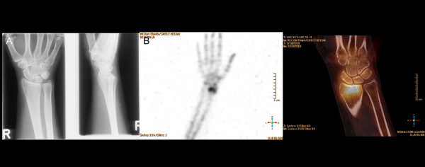
Figure 2.

A 32-year-old female with persistent dorsoradial wrist pain after distortion. (A) No bone lesion was detected on plain radiographs. (B) High radioisotope uptake in the distal radial epiphysis with bumpy RC joint surface, representing persistent bone remodeling after consolidated distal radial fracture (left: 3D-SPECT, right: fusion SPECT/CT). Occupational therapy was initiated after SPECT/CT.
