
Figure 1. HPV16-infected epidermis displays lowered and irregular E-cadherin staining HPV16 E6 expressing cells have reduced E-cadherin expression.
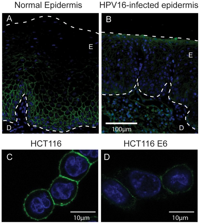
E-cadherin (green) and DAPI stained nuclei (blue) in (a) normal and (b) HPV16-infected human epidermis. E = epidermis, D = dermis. (c) HCT116 and (d) HCT116 E6 cells grown on glass coverslips, permeabilised and stained for E-cadherin (green) and DAPI to identify nuclei (blue).