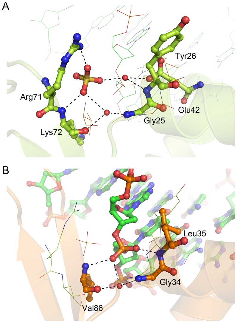
Figure 6. The bound sulfate anion in the bcPadR1 structure.
(A) Interactions between bcPadR1 residues and the sulfate ion. Also shown is the DNA-bound RTP structure (PDB entry 1F4K) onto which the bcPadR1 dimer was superimposed. The bcPadR1 residues and the sulfate ion are shown in ball-and-stick representation and labeled, while the DNA strand of the RTP-DNA complex is drawn using lines. (B) Interactions between the RTP residues and the DNA phosphate backbone adjacent to the sulfate anion in the superimposed bcPadR1 structure. The RTP residues and DNA strands are shown in ball-and-stick representation and labeled, while the bcPadR1 residues and sulfate are drawn using lines.

