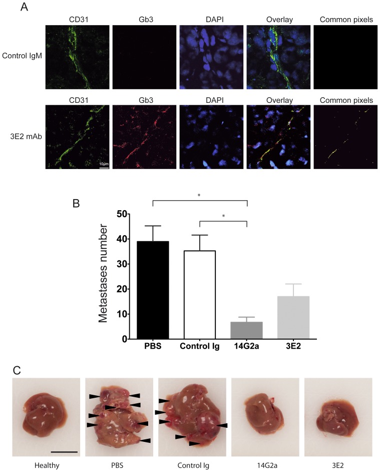
Figure 6. 3E2 inhibits in vivo metastases spreading.
A) Pictures of NXS2 hepatic metastases by confocal microscopy stained with Alexa488-conjuguated CD31 mAb (green), Alexa568-conjuguated 3E2 (red) or isotypic IgM control and counterstained with Draq5. Colocalization of the endothelial marker CD31 and Gb3 is shown by the yellow staining on the merge image. B) Number of liver metastases per animal (n = 6; mean±SEM; *p<0.05). C) Representative photography of liver 28 days after NXS2 injection and the different immunotherapies (scale bar represents 1 cm).

