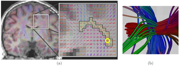
Figure 12. Estimation of  fascicles with F-test model order selection and CUSP-65.
(a) Estimated MFM superimposed on the T1-weighted anatomical image. Particularly, three tensor were correctly estimated in the centrum semiovale, which is a known brain region in which three fascicles are crossing. (b) Illustration of the tractography streamlines passing through the voxel encircled in yellow in (a), showing the three crossing fascicles.

