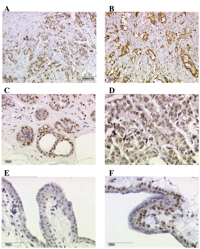
Figure 1.

Immunohistochemical staining of (A) TR, (B) RXR, (C) PPAR and (D) VDR in human breast cancer tissue. The images show immunoreactions following incubation of tumour cells with the primary antibody (×10 and ×25 lens). (E and F) Placental tissue serves as negative and positive controls for the receptors (here RXR). (E) For negative controls (blue), the isotypes matching the control antibodies of the same species were used. (F) Positive control shows TR staining of villous trophoblast cells. TR, thyroid receptor; RXR, retinoid X receptor; PPAR, peroxisome proliferator-activated receptor; VDR, vitamin D receptor.
