Figure 1.
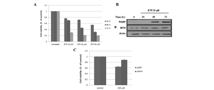
ETP-induced apoptosis in HEK293 cells. (A) ETP treatment reduced cell viability. Cells were treated with 0, 10, 25 and 50 μM ETP. After 24, 48 and 72 h, 20 μl MTT was added to each well and incubated for an additional 4 h. The results shown are the mean ± SD of 3 independent experiments. (B) ETP-induced cleavage of PARP following 48 h of incubation. Cells were incubated with 25 μM ETP for 24, 48 and 72 h. Cell lysates were subjected to SDS-PAGE. PARP and MYH expression levels were determined by western blotting using the indicated antibodies. (C) hMYH knockdown reduced cell viability following ETP treatment. Cells were transfected with siGFP or siMYH and incubated for 24 h. Cells were then treated with 25 μM ETP for 48 h. Cell viability was determined by the MTT assay. The results shown are the mean ± SD of 3 independent experiments. ETP, etoposide; IB, immunoblot; PARP, poly-ADP ribose polymerase; MYH, MutY homolog; GFP, green fluorescence protein. MTT, 3-(4,5-dimethylthiazol-2-yl)-2,5-diphenyltetrazolium bromide; SDS-PAGE, sodium dodecyl sulfate polyacrylamide gel electrophoresis; hMYH, human MYH.

