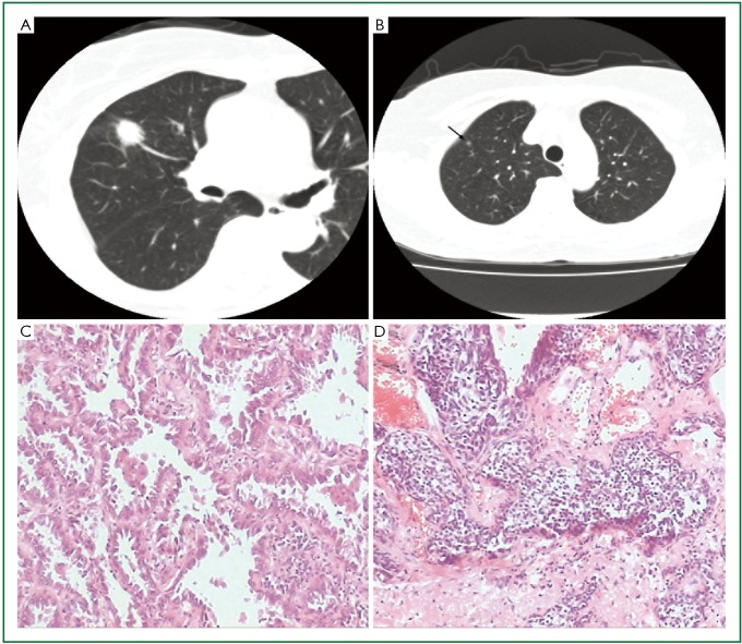
Figure 1.
A. The larger nodule was located in anterior segment; B. The smaller nodule was located in apical segment (arrow); C. The larger nodule was lung invasive adenocarcinoma with predominantly lepidic pattern and 10% papillary pattern (HE ×100); D. The smaller nodule was carcinoid tumorlet composed of a relatively uniform population cells with oval or spindle nuclei (HE ×100).

