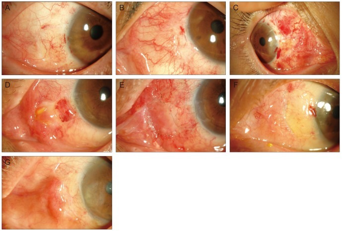
Fig. 1.
(A-C) The standard anterior segment photographs showing inflammation at 1 week after the operation (A, mild inflammation; B, moderate inflammation; C, severe inflammation). (D) Photograph of pyogenic granuloma observed in a 53-year-old woman at 1 month after she underwent pterygium excision and conjunctival autografting. (E) Photograph at 3 days after the operation when the pyogenic granuloma was removed and amniotic membrane was transplanted. (F) Photograph of dehiscence in a 57-year-old woman at 7 days after she underwent pterygium excision and conjunctival autografting. (G) Photograph of a ridge in a 71-year-old man at 28 days after he underwent pterygium excision and conjunctival autografting in his left eyes.

