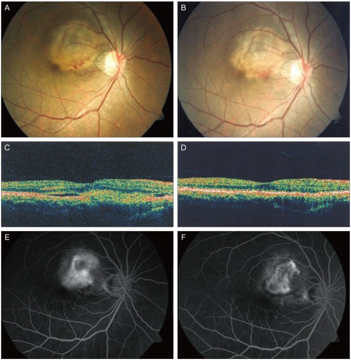
Fig. 1.
(A) Fundus photography showed a choroidal osteoma with subretinal hemorrhage, suggestive of choroidal neovascularization (CNV). (B) Fundus photography (2 weeks after treatment) showed decreased subretinal hemorrhage and decalcification of the tumor. (C) Optical coherence tomography showed the presence of CNV. (D) Optical coherence tomography (12 weeks after treatment) showed CNV. (E) Fluorecein angiography showed irregular hyperfluorecence, leakage confirmed intense CNV staining in the late stages. (F) Fluorecein angiography showed (12 weeks after treatment) that dye leakage had decreased during the late stages.

