Figure 6.

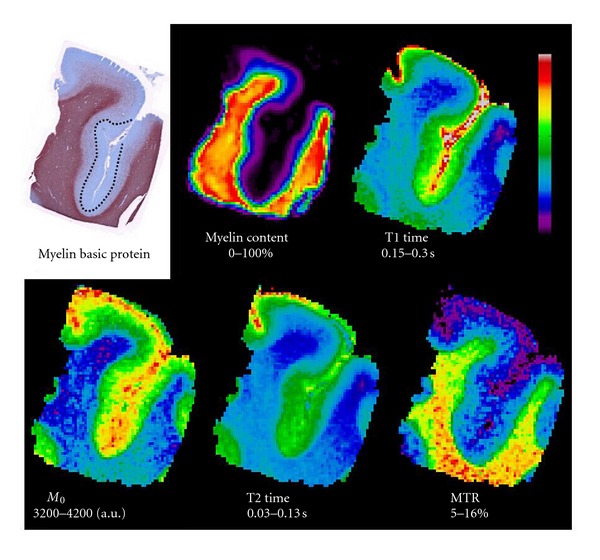
MBP stain, and co-registered myelin content and quantitative MR maps of a section of the superior frontal cortex of the MS brain with a demyelinated lesion in the fundus of the sulcus.

MBP stain, and co-registered myelin content and quantitative MR maps of a section of the superior frontal cortex of the MS brain with a demyelinated lesion in the fundus of the sulcus.