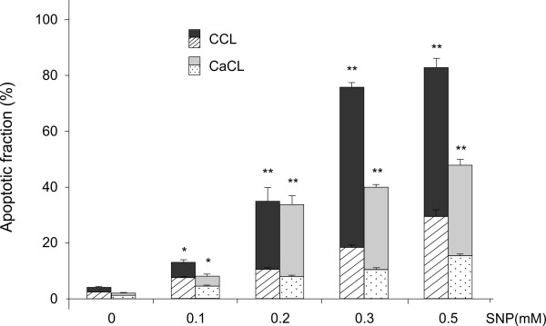
Figure 2 .
Apoptotic fraction in canine cruciate ligament cells. Canine CCL and CaCL cells were stimulated with indicated concentrations of SNP for 18 h. Apoptotic cells were measured by FITC-annexinV/propidium iodide double stained flow cytometry. The stacked bar graphs are divided into two categories: patterned graphs indicate the early apoptotic fractions detected as cells stained annexin V positive and propidium iodide negative, plain-colored graphs indicate the end stage apoptosis and death detected as cells stained annexin V and propidium iodide positive. The graphs data represent the mean ± SD from at least three separate experiments of four different cell donors, each performed in triplicates. * P < 0.05, ** P < 0.01 for combined (early and late) apoptotic fractions of CCL or CaCL treated with SNP at each indicated concentration vs. control without SNP treatment.

