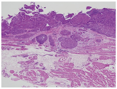
Figure 13.

pT1a-superficial muscularis mucosae case. The lower portion of the picture shows two-layered muscularis mucosae. In esophageal cancers with pT1a-MM (M3), the depth of invasion is divided into three (pT1a-superficial muscularis mucosae, pT1a-lamina propria mucosae and pT1a-deep muscularis mucosae).
