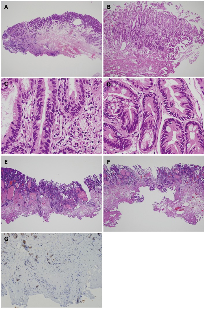
Figure 7.

Cases of positive horizontal margin and vertical margin. In most cases, a positive margin is easily recognized (A); Some cases show marked degeneration by treatment (B); By careful microscopic examination, adenocarcinoma cells (C) can be distinguished from intestinalized epithelium (D). A positive vertical margin at the submucosal layer (E) or at the lamina propria (F) should be described in the pathology report. A positive vertical margin can be easily detected by using immunohistochemistry of keratin (G).
