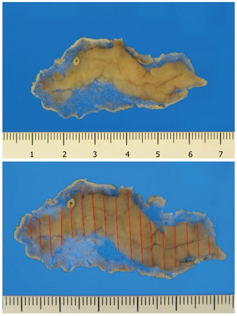
Figure 8.

Cutting of a sessile polypoid lesion. Non-tumor areas around the polypoid lesion are very thin. The submucosal layer of the tumor portion is also thin. Therefore, caution should be taken when preparing the specimen so that a false positive diagnosis of the vertical margin will not be made.
