Figure 2.

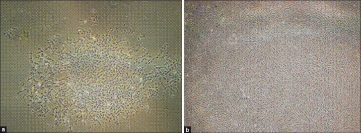
(a) Inverted microscope images of monolayer chondrocytes (a) at passage 0 after 7 days. Note the cells make colonies. (b) at passage one showing fibroblast like morphology (×60)

(a) Inverted microscope images of monolayer chondrocytes (a) at passage 0 after 7 days. Note the cells make colonies. (b) at passage one showing fibroblast like morphology (×60)