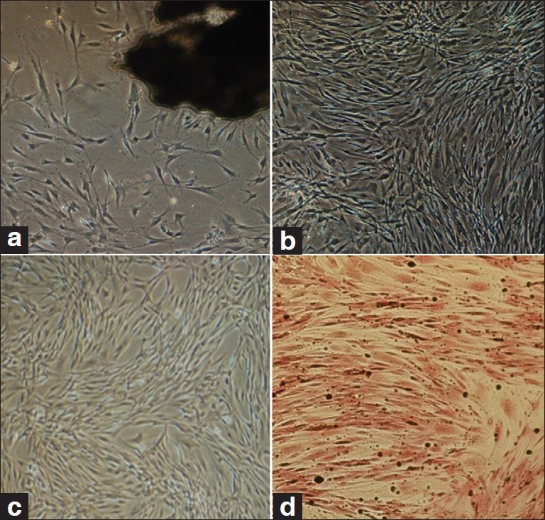
Figure 1.

(a) Explant culture of calvarial bone, cells start to isolate from bone segment (dark mass), (b) Native osteoblasts, one passage after isolation, (c) ADSCs. MSCs from third passage with spindle-shaped morphology, (d) Von Kossa staining of differentiated osteoblasts (day 14 of differentiation). Black nodules show deposition of mineralized extracellular matrix
