Figure 1.

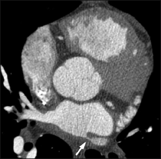
Axial CT image at the level of the left atrium demonstrates a high-grade ostial stenosis involving the left superior pulmonary vein (arrow).

Axial CT image at the level of the left atrium demonstrates a high-grade ostial stenosis involving the left superior pulmonary vein (arrow).