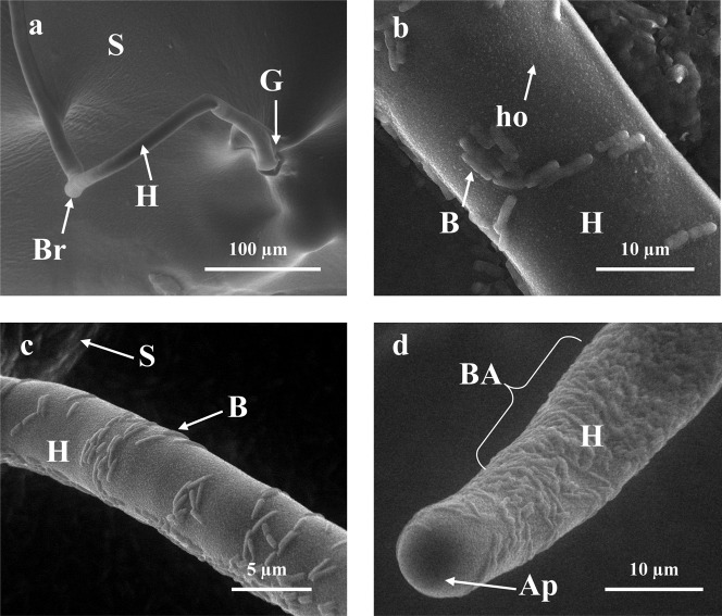
Fig. 2. In situ SEM picture of G.margarita spore (S) and hyphae (II) colonized by PE (B), including their germination (G) and branching point (Br).
Some holes (ho) and bacterial aggregates (BA) were detected on the hvphal surface, but no bacterial colonization on the apical part of hyphae (Ap). Control (no bacterial) (a): KTCIGM01 (b); KTCIGM02 (c); KTCIGM03 (d).

