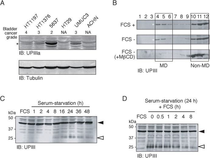
Fig. 4. Serum starvation promotes partial proteolysis of UPIIIa in 5637 cells.

(A) Expression level of UPIIIa in 5637 and some other human cancer cell lines. (B) 5637 cells were cultured in serum-containing medium (FCS +), serum-free medium in the absence (FCS −) or the presence of 15 µM MβCD (FCS −, MβCD). Cell samples were subjected to subcellular fractionation by discontinuous sucrose density gradient ultracentrifugation as in Fig. 3D, and fractions obtained were analyzed by immunoblotting with the anti-UPIII antibody. The positions of the MD and the non-MD fractions were indicated by opened squares. Asterisks indicate the positions of the 45-kDa UPIIIa. (C) Triton X-100-solubilized cell extracts were prepared from 5637 cells that had been exposed to serum-free medium for the indicated times (0–48 h). Protein samples (30 µg/lane) were analyzed by immunoblotting with anti-UPIII antibody. Closed and opened arrowheads indicate the positions of the 45-kDa UPIIIa and its degraded fragment, respectively. (D) Serum-starved 5637 cells (24 h of serum starvation) were treated with serum-containing medium for the indicated times (0–8 h). Closed and opened arrowheads indicate the positions of the 45-kDa UPIIIa and its degraded fragment, respectively. In panels C and D, control data obtained with the normally-cultured 5637 cells (FCS) are also shown.
