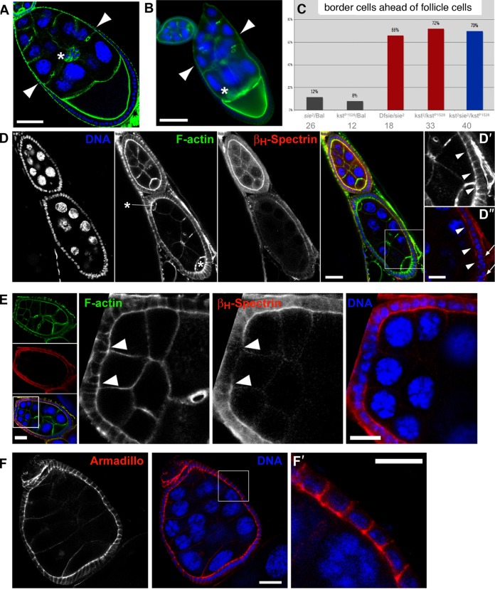
Fig. 5. Sie–kst/βH-Spectrin interaction, BC/FC migration and maintenance of epithelial structures.
(A,B) DNA is in blue and filamentous F-actin in green. Scale bars represent 50 µm. (A) Wild type stage 9 egg chamber with coordinated migration of BC (asterisk) and outer FC (arrowheads mark most anterior ones). (B) Stage 9 sie5/Df(3R)Exel6200 egg chamber with BC (asterisk) that have already reached the oocyte while the most anterior outer FC (arrowheads) have not even completed half of their migration. (C) Quantification of mis-coordination between outer FC and BC migration in heterozygous, hemizygous and compound heterozygous sosie and karst mutants. See text for details. Bal stands for balancer chromosome, which is wild type for sie and kst. Dfsie is a small deficiency that removes sie. Numbers below the genotypes indicate the number of stage 8 to 9 egg chambers counted for each genotype. Having only one functional copy of sie in a kst background did not further enhance the frequency of this phenotype. (D) Disruption of apical βH-Spectrin localization in follicle cells of a sie4/sie4 compound egg chamber. Asterisks indicate the position of the two oocytes. D′,D″ show a magnified view of the area boxed in the merge panel. Arrowheads point to the apical sides of follicle cells that have lost βH-Spectrin signal (D″), while the appearance of F-actin (D′) seems normal. Arrows in D″ point to cytoplasmic puncta of βH-Spectrin signal. Scale bars represent 20 µm in the main panels and 10 µm in D′,D″. (E) Anterior follicle cells of a sie2/sie2 compound egg chamber have lost apical βH-Spectrin signal. Left panels show an overview of the egg chamber. Note the difference in size and ploidy of the nurse cells that originated from two different cystoblasts. The boxed area denotes the region that is magnified in the panels on the right. In these, arrowheads point to the border between follicle cells that show normal apical accumulation of βH-Spectrin signal and those in which apical βH-Spectrin signal becomes virtually undetectable. Scale bars represent 20 µm in the overview panels and 10 µm in the panels showing a magnified view. (F) Adherens junctions appear normal and are precisely located in an apico-lateral position in a sie2/sie2 compound egg chamber as assayed by immunostaining for Armadillo/β-catenin. F′ shows a magnification of the area boxed in the middle panel. Scale bars represent 20 µm in F and 10 µm in F′, respectively.

