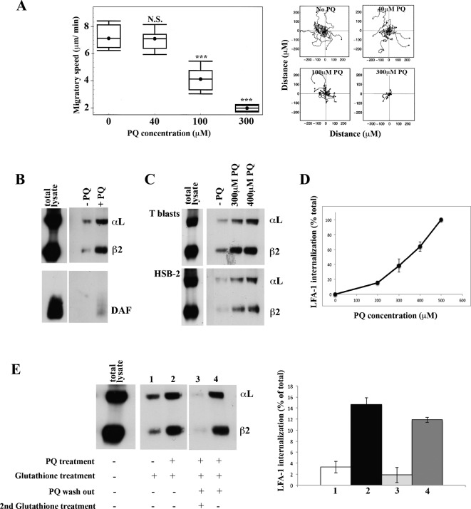
Fig. 1. LFA-1 is internalized and re-expressed by T cells migrating on ICAM-1.
(A) Speed of T lymphoblasts treated with increasing concentrations of primaquine (PQ) migrating on immobilized ICAM-1 showing cell trajectories tracked by video microscopy. Mean speed ± s.e.m. of three independent experiments (left) and the migratory tracks in a typical experiment (right) are shown; n = 20 cells per condition, ***P<0.001. (B) Western blots of immunoprecipitated internalized LFA-1 and DAF following biotinylation of T lymphoblast surface membranes followed by 30 min migration on ICAM-1: total cell lysate immunoprecipitated for LFA-1 and DAF (sample diluted 2×) and internalized protein ± 300 µM PQ revealed by removal of biotin from cell membrane receptors with glutathione. (C) Western blots comparing internalized LFA-1 in T lymphoblasts and HSB2 T cell line: total biotinylated LFA-1 (total lysate at 2× dilution) and similar amounts of internalized LFA-1 ± PQ. (D) Internalized T cell LFA-1 after 30 min on ICAM-1 in the presence of increasing amounts of PQ ± s.d. from n = 3 experiments. (E) Total biotinylated LFA-1 (total lysate at 2× dilution). Lanes 1 and 2: LFA-1 internalized after 30 min ± 300 µM PQ. LFA-1 re-exposure on the membrane following PQ washout is demonstrated by lack of intracellular LFA-1 in lane 3 (treated with glutathione to remove detection of membrane LFA-1) and lane 4 (no glutathione treatment allowing membrane and intracellular LFA-1 to be detected). Left: typical experiment. Right: mean ± s.d. of n = 3 experiments.

