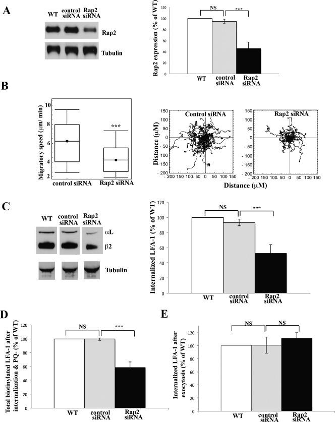
Fig. 2. Rap2 regulates internalization of T cell LFA-1 during migration.
(A) HSB2 T cells were either not electroporated as control WT or electroporated with control or Rap2 siRNAs. Western blots were probed for Rap2 after 72 h and α-tubulin as a loading sample control. Left: typical experiment showing Rap2 siRNA knockdown compared with non-electroporated (WT) or control siRNA-treated T cell controls. Right: quantification of siRNA knockdown compared with controls, mean ± s.d. of n = 5 experiments, ***P<0.001. (B) HSB2 T cells electroporated with control or Rap2 siRNAs migrating on ICAM-1 for 30 min. Left: mean speed ± s.d. of n = 3 experiments. Right: migratory tracks of individual cells; n = 40 cells per condition, ***P<0.001. (C) Biotinylated LFA-1 internalization + PQ in WT, control siRNA- and Rap2 siRNA-treated HSB2 T cells. LFA-1 internalization following Rap2 siRNA compared with controls, showing (left) Western blot of a typical experiment and (right) quantification of 3 experiments, mean ± s.d. ***P<0.001. (D) Using T cells treated as in C, total biotinylated LFA-1 in control and Rap2 siRNA-treated T cells is shown following PQ washout and a 20 min incubation. No glutathione treatment allows assessment of internal and re-exposed LFA-1; mean ± s.d. of 3 experiments, ***P<0.001. (E) Re-expression of LFA-1 in control and Rap2 siRNA-treated T cells following PQ washout and 20 min incubation. LFA-1 remaining inside the cells revealed following removal of glutathione sensitive membrane LFA-1; mean ± s.d. of 3 experiments, ***P<0.001.

