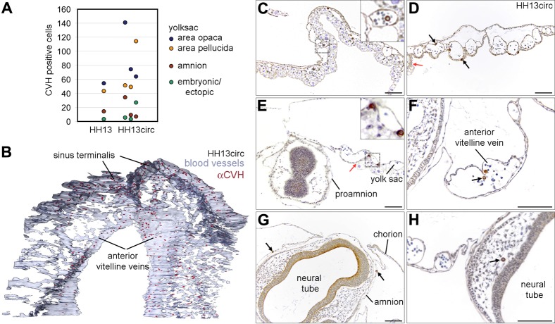
Fig. 3. At HH13circ the majority of PGCs is localized in the sinus terminalis and anterior vitelline veins.
(A) Analysis of position of PGCs in sectioned embryos at stage HH13circ. PGCs at HH13 and HH13circ are present in similar numbers in the yolk sac in the anterior area opaca and pellucida; the amnion and ectopically in the embryo head. (B) 3D reconstruction of the extraembryonic vasculature of embryos at HH13circ has shown that the PGCs were mainly localized in the anterior vitelline veins and the sinus terminalis. (C–E) Transverse sections of HH13circ embryos immunostained for CVH. PGCs were dispersed in the area opaca (C) and area pellucida (D) anterior from the head and at the level of the head (E). PGCs were observed inside and outside the blood vessels (black arrows). The junction between the area opaca and pellucida is marked by a red arrow. Note in E, that the head at the level of the prosencephalon is completely surrounded by proamnion. (F) PGCs (black arrow) in the anterior anterior vitelline veins. (G,H) Ectopic PGCs (black arrows) were found in the amnion (G) and in the capillary network of the head (H). Scale bars: 100 µm in C–H.

