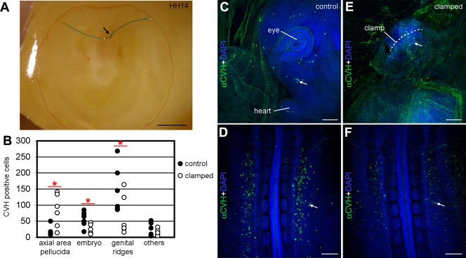
Fig. 4. Blocking the anterior vitelline veins prevented the correct migration of PGCs towards the genital ridges.
(A) The anterior vitelline veins were clamped in HH14 embryos growing in ovo and the embryos were allowed to develop for 6 hours. (B) Analysis of the total number of PGCs in control (n = 7, black dots) and experimental embryos (n = 6, white dots) in different regions. The differences in distribution of the PGCs in the axial area pellucida, the embryo and genital ridges were statistically significant (P<0.05) using the non-parametric Mann–Whitney test [(*) P<0.05]. (C–F) The number of PGCs (white arrows) present ectopically in the embryo head (C) and genital ridges (D) was consistently higher in control embryos than in experimental embryos, where the PGCs concentrated surrounding the clamped vitelline veins (E) and the number of PGCs settled in the genital ridges was reduced (F). Scale bars: 500 µm in A, 100 µm in C,E and 200 µm in D,F.

