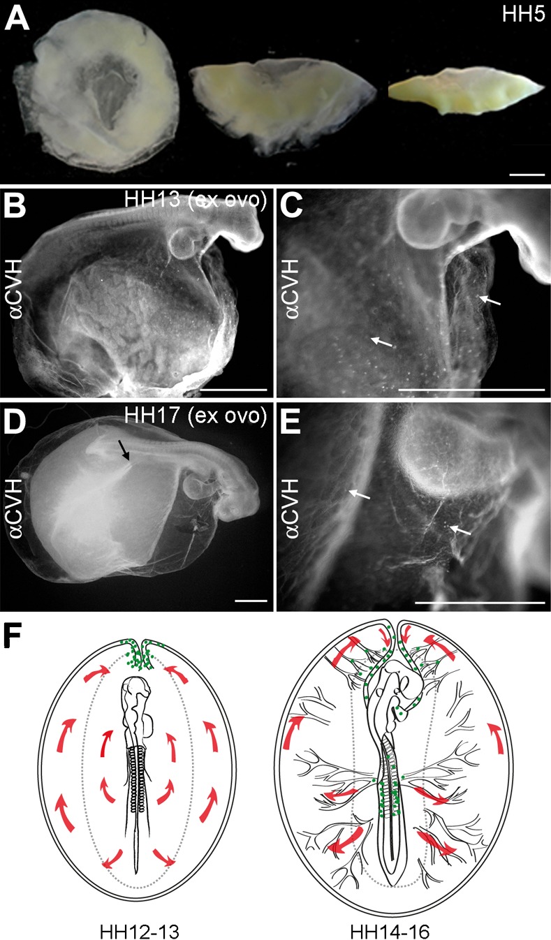
Fig. 5. A new model for PGCs migration in chicken embryos.

(A) Embryos at HH5 prepared to be cultured using the Cornish pasty method. (B,C) Ex ovo embryos at HH13 showed a relatively normal embryonic morphology (B) and the PGCs were observed in the germinal crescent area in both the somatopleura and splanchnopleura (white arrows). (D,E) Ex ovo embryos at HH17 showed a relatively normal embryonic morphology and the formation of well-developed posterior vitelline arteries (black arrow) (D) and the PGCs were still observed in the germinal crescent area in both the somatopleura and splanchnopleura (white arrows) (E). (F) A new model for PGCs migration in chicken embryos. At HH12–13, the yolk sac circulation courses in loop (red arrows) to enter the embryo via the heart. At this stage, the majority of PGCs (green dots) localized axially at the border between the area opaca and pellucida, where the sinus terminalis converged in the anterior vitelline veins. At HH14–16, the PGCs (green dots) circulated effectively towards the embryo via the sinus terminalis and the anterior vitelline veins towards the heart. Thereafter, the PGCs traffic via the aorta to the caudal part of the embryo and become lodged in the genital ridges. Scale bars: 100 µm in A–E.
