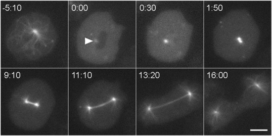
Fig. 1. Mitosis in Dictyostelium.

Still frames from a live cell recording, showing GFP-tubulin distribution as a cell transitions from interphase (−5:10) into mitosis and continues into cytokinesis. Although this cell lacks the Kif9 protein, it shows the process and timing characteristic of wild-type cells. The interphase microtubule array rapidly disassembles at the G2/M transition, leaving a dim spot to mark centrosome position (arrowhead, 0:00) adjacent to the dark area of the nucleus. At 30 s, the nucleus has flooded with tubulin. The centrosome is noticeably brighter indicating that it has docked into the nuclear envelope and begun to incorporate tubulin into a nascent spindle. At 1:50, the replicated centrosomes have separated into a bipolar, prometaphase configuration. The subsequent three frames show spindle elongation. In the final frame, the spindle has disassembled, astral microtubules have grown, and the cleavage furrow is constricting the cell into two. Time in min:sec, bar = 5 µm.
