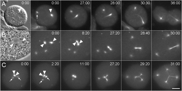
Fig. 7. Daughter centrosome dynamics.
Three sequences that demonstrate spindle arrangements resulting from daughter centrosome–nuclear interactions. (A) Mononucleated kif9 null cell with a single centrosome. Daughter centrosomes have already separated at 0:00: the significantly brighter daughter (arrowhead) has docked into the nuclear envelope while other daughter (arrow) remains in the cytosol. The first frame shows a DIC image where the nucleus is clearly visible; the subsequent frames show GFP-tubulin fluorescence at the times indicated. The single nuclear daughter forms a monopolar spindle as this cell progresses through mitosis. (B) Mononucleated kif9 null cell with two centrosomes. Both centrosomes have separated forming four daughters; two of which have independently docked into the nuclear envelope (0:00) (arrowheads). The first frame shows a phase contrast image where the nuclear daughter centrosome positions are indicated around the relatively clear area of the nucleus. The two nuclear daughter centrosomes coalesce to form a functional bipolar spindle apparatus, demonstrating plasticity of the spindle assembly process. (C) Mononucleated kif9 null cell with two centrosomes. One centrosome has docked into the nucleus and has split into a prometaphase spindle arrangement (arrowheads in 0:00), while the other centrosome has separated into daughters in the cytosol (arrows). By 2:10, one of the cytosolic daughter centrosomes has docked into the nucleus (right arrow) and has begun to incorporate GFP-tubulin. The three nuclear daughter centrosomes then proceed to form a tripolar spindle apparatus, while the unengaged daughter remains in the cytosol. Time in min:sec, bar = 2 µm.

