
Fig. 2. Taperin is predominantly nucleoplasmic in vivo.
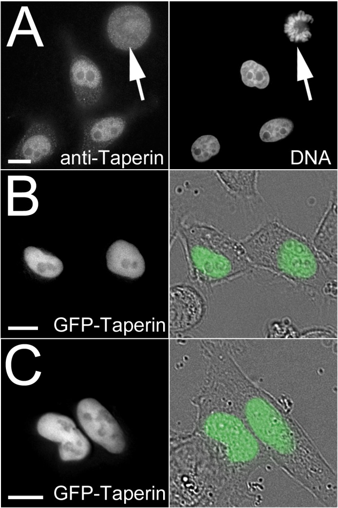
(A) Affinity purified taperin antibodies (5 µg/mL) were used to localize endogenous protein and DNA was stained with Hoechst in PFA-fixed HeLa cells (top panel), revealing a predominant nucleoplasmic localization in interphase and diffuse staining with no apparent accumulations in metaphase (arrow). Taperin is also predominantly nucleoplasmic when transiently expressed as a GFP-tagged fusion protein in both HeLa (B) and U2OS (C) cells. In these two panels the GFP images are shown superimposed on differential interference contrast (DIC) images to the right. Scale bars are 5 µM. Antibody controls are shown in supplementary material Fig. S3.