
Fig. 3. Cx26 and Cx32 form heteromeric connexons in Xenopus embryos.
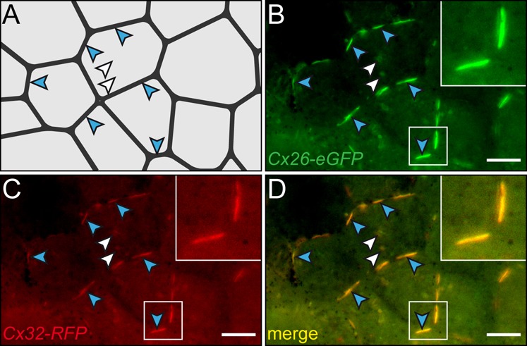
Fusion constructs (Cx26-eGFP and Cx32-RFP) were co-injected into the animal region of single blastomeres at the 4-8 cell stage and cultured through blastula-gastrula stages. Fluorescent fusion proteins were co-expressed in ectodermal cells of the animal cap region. Expression was found in vesicles (white arrowheads) and the plasma membrane (blue arrowheads). (A) Cell boundaries. Positions highlighted in (B–D) are indicated by arrowheads. (B) Cx26-eGFP staining. (C) Cx32-RFP staining. (D) Overlay of frames. Scale bar represents 10 µm.