
Fig. 5. Comparable expression of epicardial genes in regenerating hearts of young and old fish.
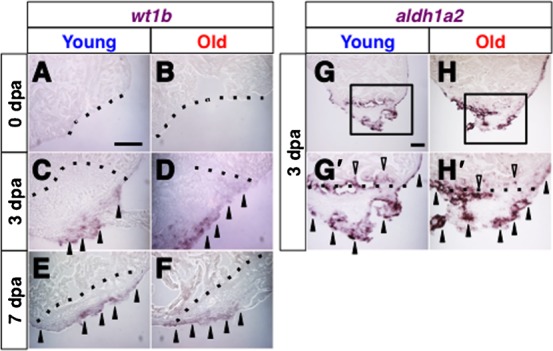
(A–F) In situ hybridization of wt1b immediately after amputation at 0 dpa (A,B), at 3 dpa (C,D) and at 7 dpa (E,F) in regenerating hearts of young (A,C,E) and old (B,D,F) fish. Arrowheads point to the wt1b signals. (G–H′) in situ hybridization of aldh1a2 at 3 dpa in the regenerating hearts of young (G,G′) and old (H,H′) fish. Black arrowheads and open arrowheads point to the aldh1a2 signals in the epicardial tissue and endocardial tissue, respectively. G′ and H′ shows close up images of the boxed areas in G and H. Dotted lines indicate the amputated planes. Scale bar: 50 µm; the degree of zoom: ×2.